-40%
USA Digital Dental Clinic X-Ray RVG Sensor Portable X-Ray Machine Image Sensor
$ 47.51
- Description
- Size Guide
Description
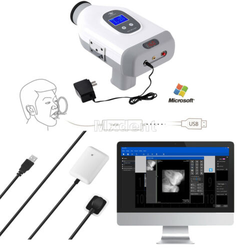
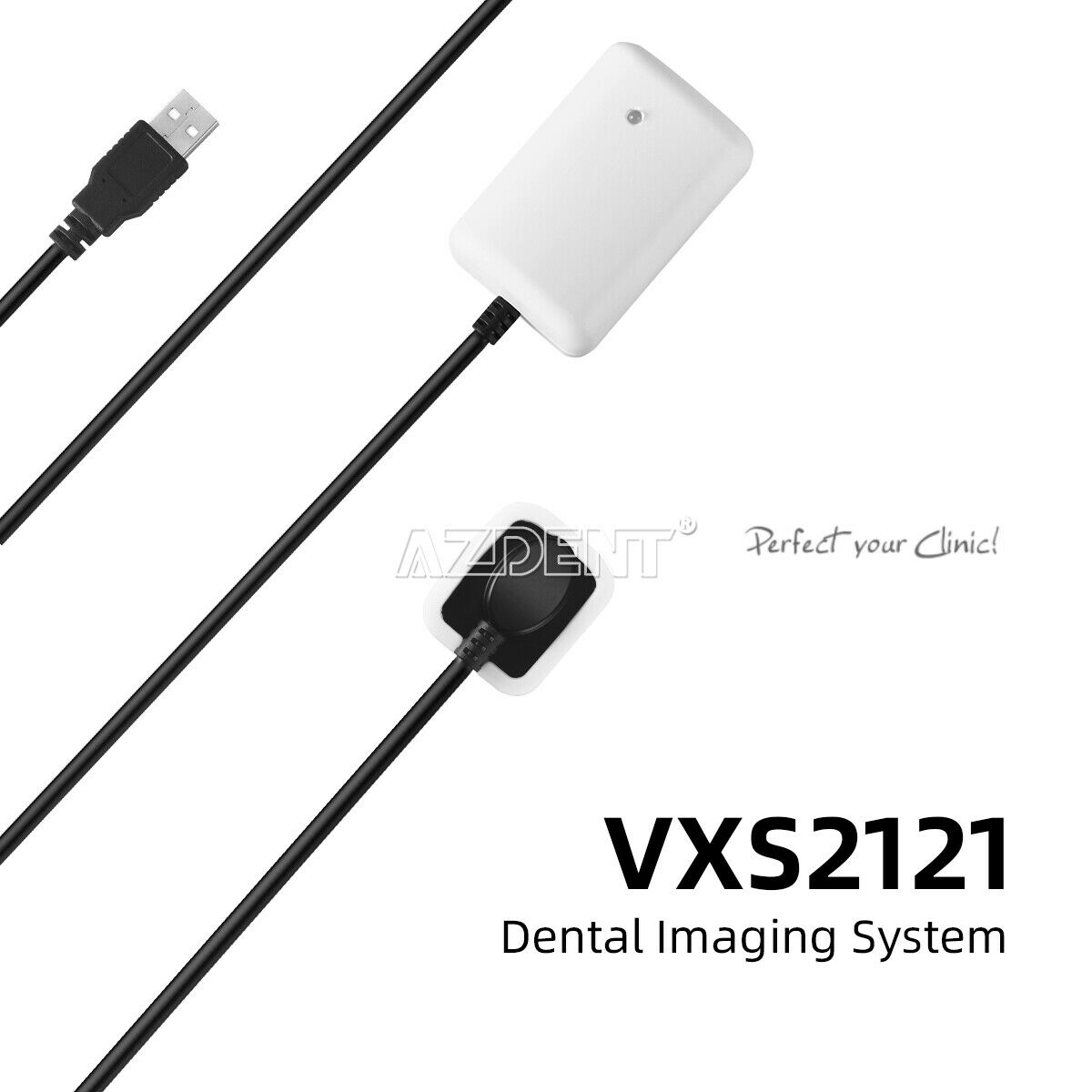
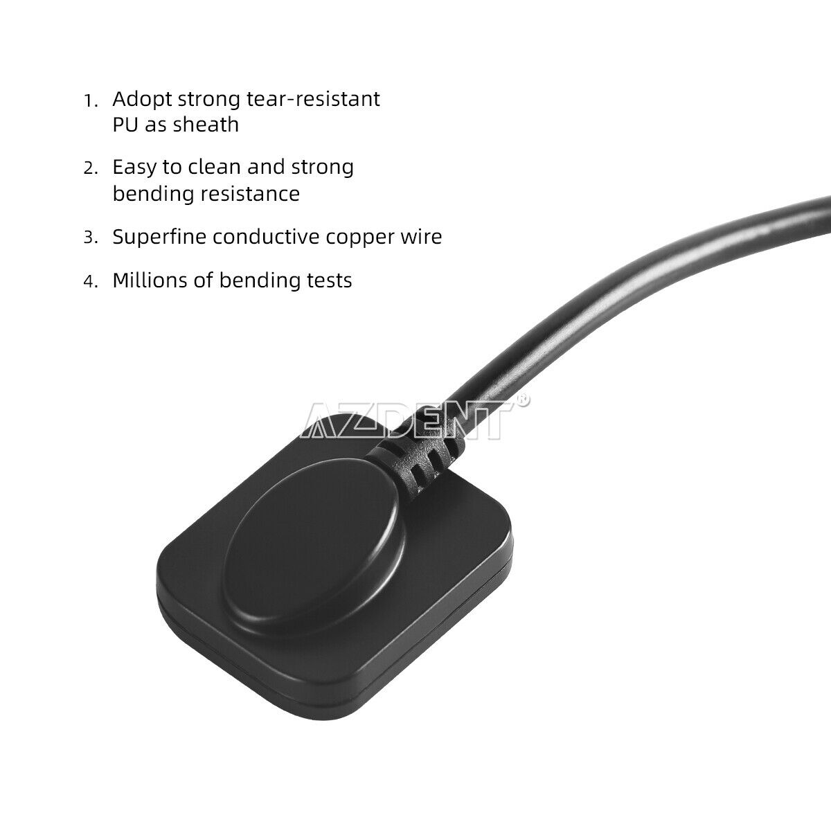
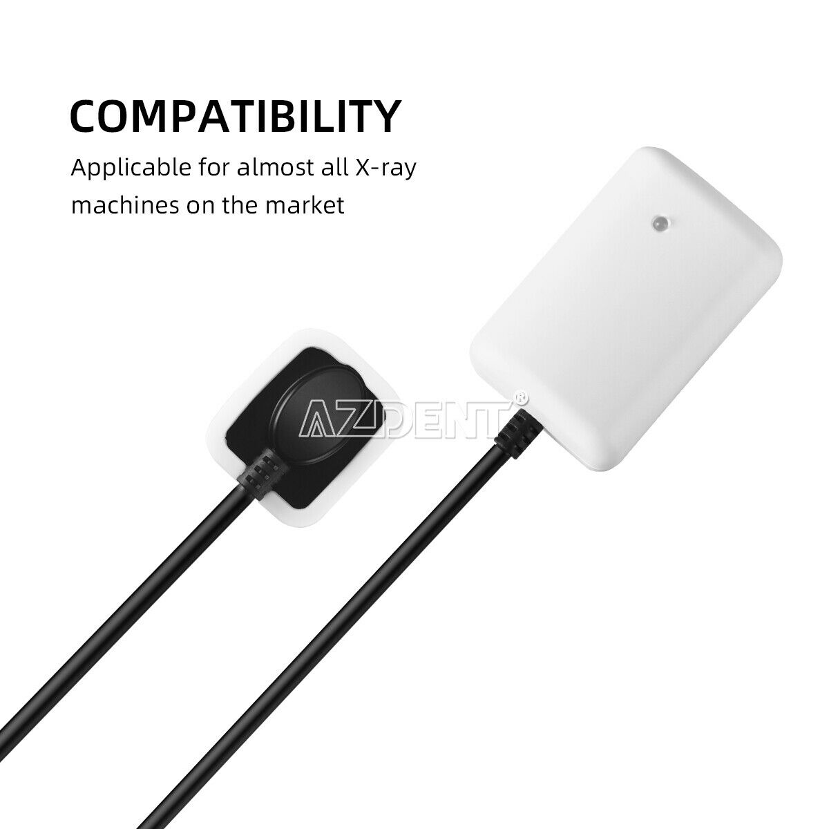
Features:The product is mainly composed of a sensor, an adaptor, a USB connection cable and workstation software. Working together with an X-ray equipment, the Product is intended to make X-ray radiography in dental clinics or veterinary clinics,Sensor size Suitable for child(adult single tooth)and pets.
Hardware Requirements:
Central Processor: Intel Core i3 or higher
Internal Memory: 4GB or higher
Hard Disk: 80GB or higher
Display: 1280X1024
USB interface: two
Keyboard: One
Mouse: One
Software Requirement
Operating Software:Windows 7 SP1,8,10 32/64bit
Runtime: .Net Framework 4.6 or higher, VC++ 2015
Specification:
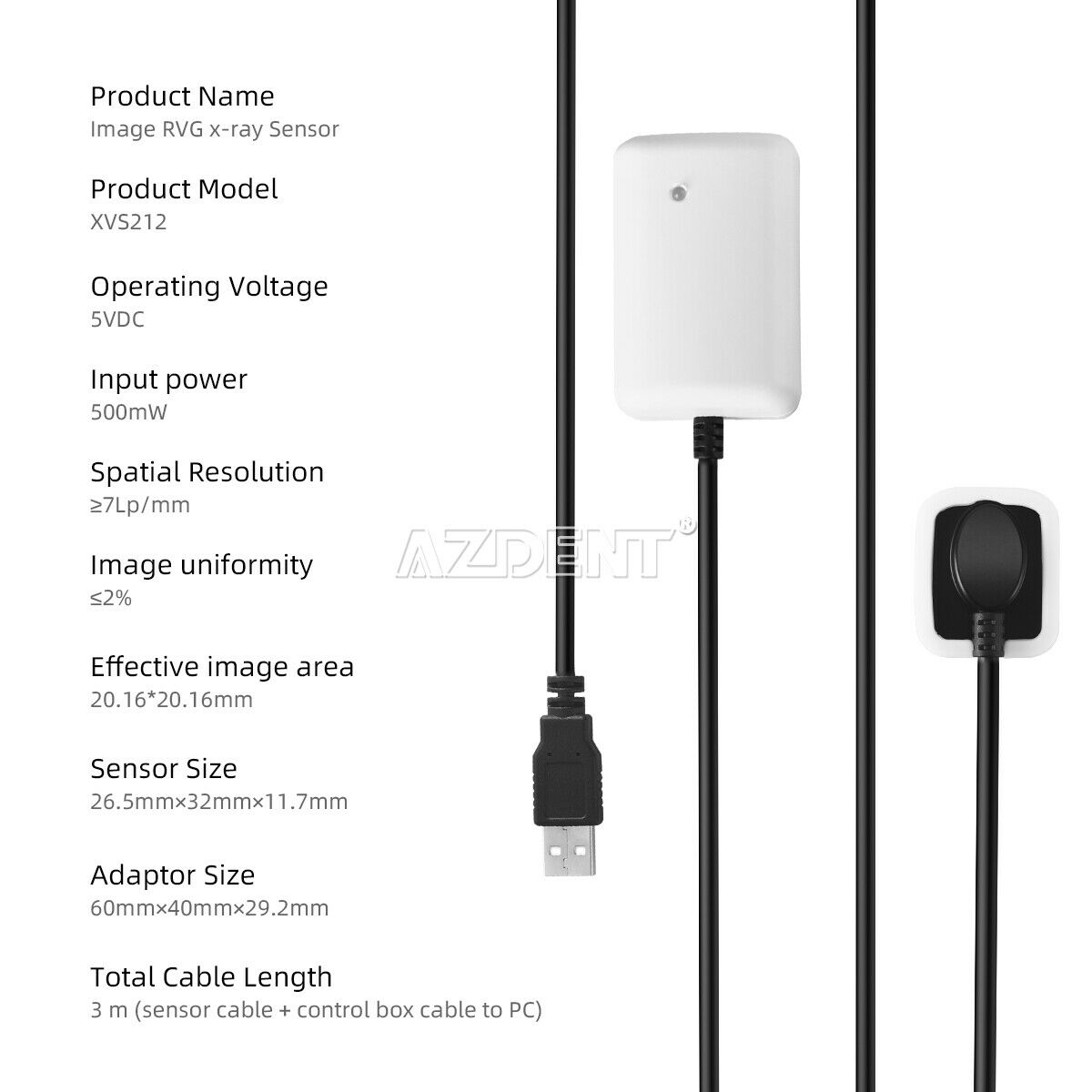
Product Name: Image RVG x-ray Sensor
Product Model:XVS2121
Operating Voltage:5VDC
Input power:500mW
Spatial Resolution:≥7Lp/mm
Low contrast resolution :Able to display holes with 1mm, 1.5mm, 2mm and
2.5mm diameter on the 0.5mm-thick aluminium foil
Image uniformity ≤2%
Effective image area: 20.16*20.16mm
Sensor Size:26.5mm×32mm×11.7mm
Adaptor Size:60mm×40mm×29.2mm
Total Cable Length: 3 m (sensor cable + control box cable to PC)
Warranty:1 Year
Accessory :
Digital sensor: 1 Set
USB cable :1 PCS
U Disk: 1PCS
Disposable protective sheath: 1 Box(500)
silicon loop: 1 PCS
Sensor holder: 1 PCS
Aluminum plate:1 PCS (40*40*6mm)
++
++++++++++++++++++++++++++++++++++++++++++++++++++++++++++++++++++++++++++
Product introduction:
This model combines the advantages of similar products found at home and abroad; it eliminates the shortcomings of the on-frequency X-ray machine (high current intensity and excess amounts of scrap x-rays).Using a Toshiba 0.3 micro-focus tube.The tube voltage frequency of this machine is 30 KHz
,
and the tube current is 1 MA. The radiation scope is at an angle of 24 degrees
,
located within a distance of 1.2 meters ahead. This is an innovative Chinese design named the "Green X-ray Machine."
Warning: After turning on the machine, wait for 1 minute, the voltage can be stabilized before normal use
;
After each photo, you have to wait 30 seconds before you can take a normal photo
。
TECHNICAL SPECIFICATIONS
:
Tube Voltage
65kv
Tube Current
1mA
Time Exposure
0.1~~1.8s
High pressure generator
30kHz DC
Rated Power
60w
Focal Spot To Skin Distance
>20CM
Focus Spot Size
0.3mm(TOSHIBA)
Charger Input Voltage
AC110V
~
220V
Supply Frequency
30Hz
Radiation Leakage
<20ugy/h
Battery
24V DC 2600MA
Weight
4.3KG
Dimensions
37 cm×31 cm×28cm (L×W×H)
Reference of the Angles
:
'
Tooth
Upper
Lower
1-2
42
-15
3
45
-18
4-5
30
-10
6-8
28
-5
Reference of the time of exposure
:
Tooth
Adult
Child
Central & Lateral
0.2-0.3
s
0.2-0.3
s
Cuspid & 1st Bicuspid
0.3-0.4
s
0.3-0.4
s
Upper 5
,
6
0.5-0.6
s
0.5-0.6
s
Upper 7
,
8
1.3-1.8
s
1.2-1.5.
s
Lower 5
,
6
0.5-0.6
s
0.4-0.5
s
Lower 7
,
8
0.6-0.8
s
0.5-0.7
s
Packing List:
Main machine
1pcs
Power line
1pcs
User manual
1pcs
Operation of the Main Unit :
Upon receiving
,
open the box and check the product for possible damage during shipping.
Make sure the fittings on the encasement list are packaged within the box.
Turn on the Power
;
the pilot lamp will be light up.During this time
,
the digital tube
will show the fore setting time. Now the equipment is in standby mode.
Setting the time (Skip to next step if unneeded)
→
then press "+" and" -" to set the time needed (time range is 0.1-1.8seconds); The equipment is now in standby mode.
Put the Tooth film plumb behind the tooth which is going to be taken picture
,
and be as close as possible (the smooth side stick to the tooth)
Keep the ball head plumb to the tooth projection position; Have the ball head
,
tooth
,
and tooth film steadily mutually plumb.
After positioning
,
use the ON/OFF on the main Unit
,
to take pictures. (Notice: gently press 0.5 sec to turn it on)
Points for Attention
:
Make sure the angle of film
,
tooth
,
and ball head are properly aligned when taking a picture. Keep them steady until the last step of picturing; making sure that there is no change in any of their positions.
Remember to turn the power 0ff when work is done.
Charge the battery if the machine cannot work and the red lamp lignt
After turning the power on
,
wait one minute before taking pictures
,
this allows the ray to provide a steady output.
The equipment switches into protection mode automatically when the voltage is incorrect. If this occurs
,
it will not be able to carry out the normal work of filming.
When taking pictures
,
the ON/OFF button could stop the objection and then the machine goes back to the preparation mode.
Use high quality tooth film and developing liquid to make clear pictures.
Handle the ball head gently while in use
,
so as not to damage the delicate component.
If a situation occurs
,
preventing the machine from taking regular pictures
,
contact the seller to solve the problem instead of trying to service the product yourself.
Keep the tooth film and the liquid in a proper place and use them within the shelf-life period.
Development of films should be performed at temperatures ranging between 23-25 degrees Celsius to ensure the image quality.
Other requests accordance with the scope of the technical parameters.
Please read the manual carefully before using.
FDA Statement
The sale of this item may be subject to regulation by the U.S. Food and Drug Administration and state and local regulatory agencies. If so, you can bid on this item only if you are an authorized purchaser. If the item is subject to FDA regulation, I will verify your status as an authorized purchaser of this item before shipping of the item.
Seller name:MXDENT
City,State:SHENZHEN,China
Telephone number:0755-36925817
System, X-Ray, Mobile
K200976
SR-8230, SR-8230S Portable X-Ray Unit
892.172
IZL
4/13/2020
6/10/2020
Substantially Equivalent (SESE)
Radiology
Radiology
Summary
Traditional
No
No
